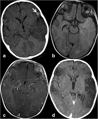
Fig. 3.

A 7-month-old infant with herpes encephalitis. At presentation (a), asymmetric hypodensity is noted in the left temporal lobe on noncontrast CT and subtle hypodensity in the right temporal lobe. Hemorrhagic focus is noted on the left frontal operculum (arrow). MRI at presentation shows bilateral temporal lobe FLAIR hyperintensity left greater than right (b) and leptomeningeal enhancement (c). Delayed contrast-enhanced CT (d) shows development of encephalomalacic changes in the temporal lobes bilaterally, left greater than right and in the right frontal lobe
