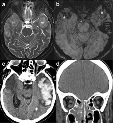
Fig. 5.

Mucormycosis infection of the temporal lobes bilaterally in a 62-year-old patient, immuncomprmised due to steroid treatment. Increased T2 signal (a) and blood products (b, SWI) are noted in the anterolateral temporal lobes (asterisks). The patient later developed massive left temporal lobar haemorrhages (c), and deceased. On coronal CT image of the same patient (d), note partial opacification of the ethmoid and maxillary sinuses and stranding of the intra-orbital fat. Autopsy revealed mucormycosis in brain parenchyma and paranasal sinuses
