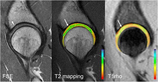
Fig. 10.

Sagittal PD-weighted MRI of the hip in a 30-year-old woman demonstrates mild chondral hyperintensity over the anterosuperior acetabular dome (arrowhead), with corresponding prolongation of relaxation times on T2 mapping and T1rho images (white arrows)
