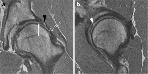
Fig. 6.

A 26-year-old man with FAI. a Coronal PD-weighted image shows a cam lesion at the femoral head–neck junction (open arrow). There is chondrolabral separation, with a cleft between the labrum and cartilage (arrow). A paralabral cyst is also seen (black arrowhead). b Sagittal PD-weighted image shows chondral delamination near the transition zone anterosuperiorly (white arrowhead)
