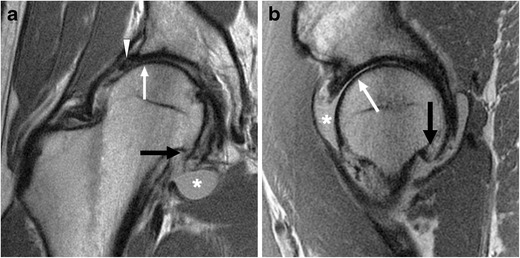
Fig. 9.

A 36-year-old man with cam-type FAI and severe osteoarthritis. Coronal (a) and sagittal (b) PD-weighted MR images of the right hip demonstrate chronic degeneration of the labrum (arrowhead). Chondral loss with extensive bone-on-bone contact is seen over the superior femoral head (white arrow). Subcapital femoral neck osteophytes are also seen (black arrow). Reactive synovitis with effusion is demonstrated (asterisk)
