
Fig. 1.
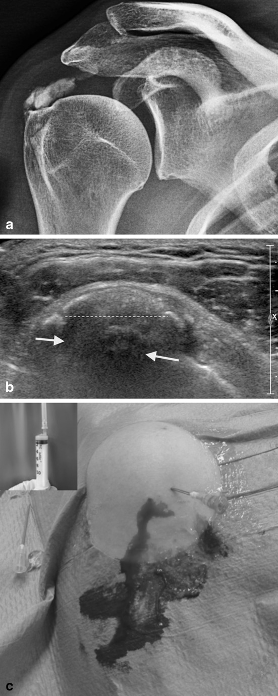
A case with acute calcifying tendinitis of the rotator cuff. (a) X-ray shows a large calcium deposit (>1.5 cm) at the insertion of the supraspinatus tendon in touch with the greater tuberosity; (b) ultrasound image in the same patient as a demonstrates a large fragmented and punctate calcification (dotted line) with hypoechoic area indicating oedema associated with the reabsorptive phase (white arrows); (c) ultrasound-guided needling and lavage in the same case as a and b with an abundant leakage of calcium (the window on the left shows the calcium aspirated in a syringe)