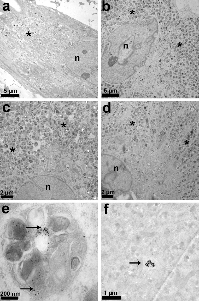
Fig. 5.
TEM analysis at day 21: a control, b Mg2Ag, c Mg10Gd, d WE43, e lysosome of HRD cultured with Mg2Ag (note the degradation particles, arrows); f cytoplasm of HRD cultured with Mg2Ag (note the degradation particles, arrows). Asterisk lysosomes, endocytotic vesicles, n nucleus. Note the high amount of lysosomes and endocytotic vesicles in b–d

