Fig. 3.
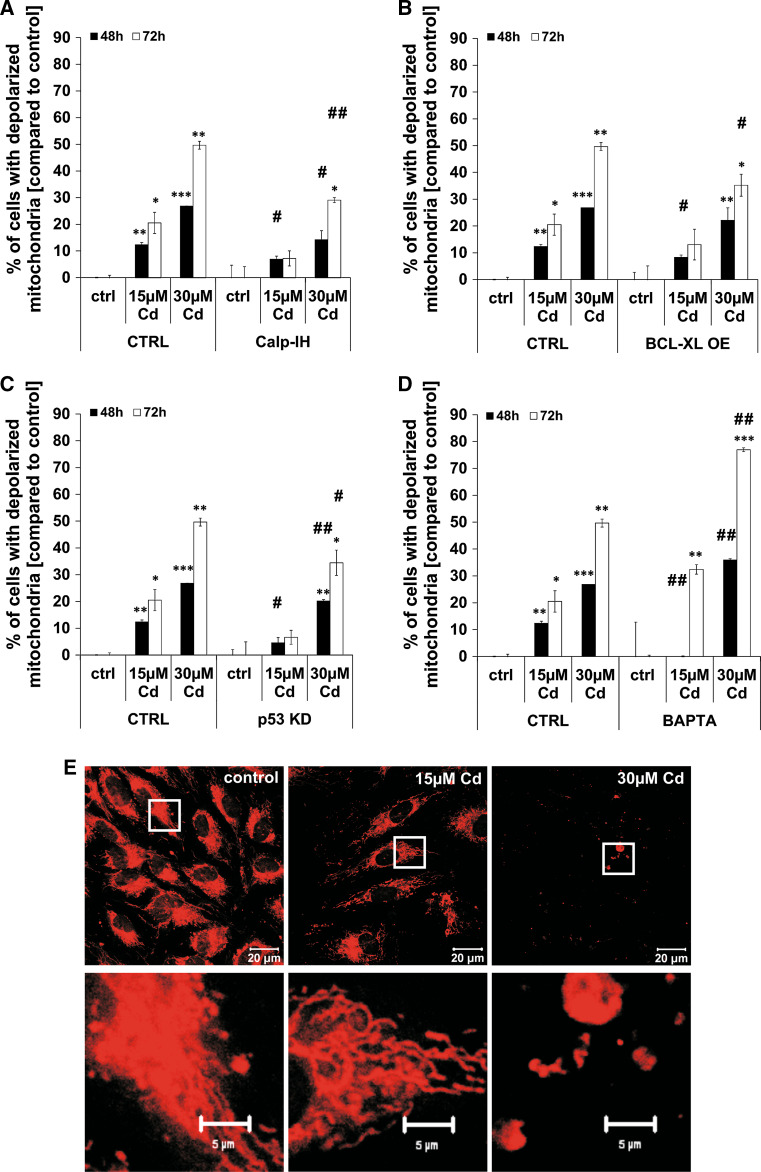
Cd impairs mitochondrial membrane potential and reduces mitochondrial mass. Flow cytometry-based analyses of mitochondrial membrane potential in Cd-treated endothelial cells are shown in (a–d). Cd-treated endothelial cells were incubated either with the calpain inhibitor or BAPTA and mitochondrial membrane potential was assayed using a JC-1 assay and quantified by flow cytometry analyses (a, d). b, c The effects of either p53 KD or BCL-XL OE on mitochondrial membrane potential in Cd-treated endothelial cells. e The first row shows representative images of Cd-treated endothelial cells (48 h) stained with MitoTracker Red and the second row shows magnifications indicated by the corresponding white boxes. All experiments were performed in triplicates and were repeated at least three times. Results depict the mean ± standard deviation. Asterisks indicate significant differences compared to the corresponding (*p < 0.05; **p < 0.01, ***p < 0.001) and hash signs indicate significant differences between the groups (# p < 0.05; ## p < 0.01)

