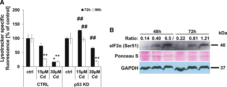
Fig. 4.
Cd-induced lysosomal membrane permeabilization and eIF2 alpha phosphorylation. Flow cytometry-based quantification of LMP was performed in endothelial cells treated with 15 and 30 µM Cd for 72–96 h. The effect of the p53 KD on Cd-induced LMP is depicted in (a). Western Blot-based analyses of eIF2 alpha phosphorylation in endothelial cells after Cd treatment is depicted (b). To compensate unequal protein loading, the Ponceau staining of membranes as well as the GAPDH expression is shown and the ratio between specific signal intensity and protein loading (GAPDH) was calculated and depicted. All experiments were performed in triplicates and were repeated at least three times. Results depict the mean ± standard deviation. Asterisks indicate significant differences compared to the corresponding control (*p < 0.05; **p < 0.01) and hash signs indicate significant differences between the groups (## p < 0.01)

