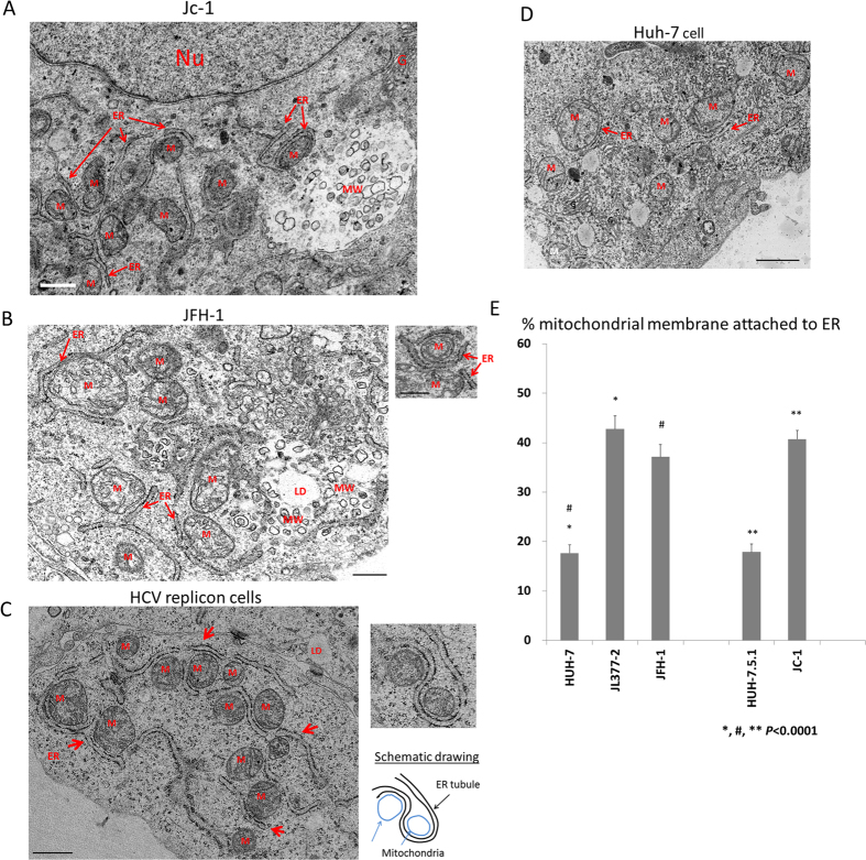
Figure 1. Extensive attachment of ER tubules to mitochondria in HCV replicon cells JL377-2 and hepatocytes transfected with JFH-1 or infected with HCVcc.
(A) Electron micrographs of Huh-7.5.1 cells infected with HCVcc. Scale bar = 0.5 μm. (B) EM images of Huh-7 cells transfected with JFH-1 full-length HCV genomic RNA. Scale bar = 0.5 μm. (C) EM images of the cytoplasm of a JL377-2 cell. Scale bar = 1μm. (D) EM image of naïve Huh-7 cells (left panel). Scale bar = 1μm. Abbreviations: M, mitochondria; ER, endoplasmic reticulum; G, Golgi; Nu, nucleus; MW, membranous web, LD, lipid droplet. (E) Quantification of the amount of mitochondria membrane tethered with ER. The percentage of perimeter membrane of mitochondria closely apposed to ER tubules in EM images of the indicated HCV harboring cells or naïve Huh-7 or Huh-7.5.1 cells were quantified. N = 60; error bars = S.E.M.; p < 0.0001.

