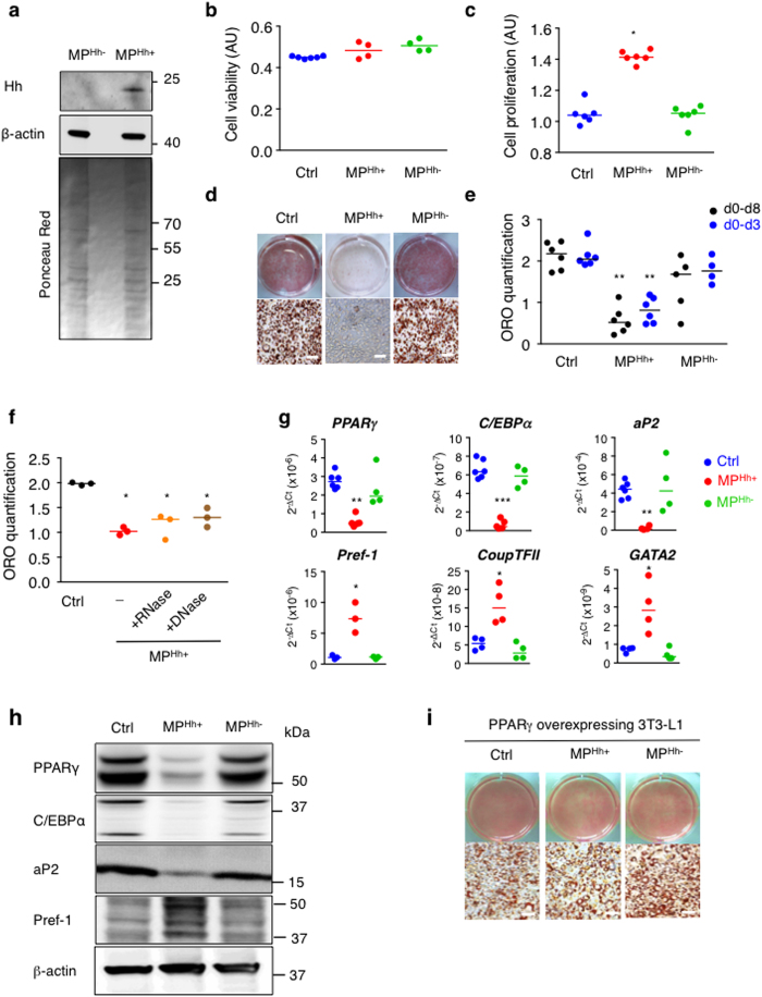
Figure 1. MPHh+ stimulate proliferation and inhibit 3T3-L1 adipocyte differentiation.
(a) Immunoblot showing specific association of Hh with MP isolated from supernatants of PMA/PHA/ActD-stimulated CEM-T lymphocytes (MPHh+) compared to ActD-stimulated CEM-T cells (MPHh−). β-actin, known to associate with MP, is used as a loading control. Ponceau red staining of the membrane shows equal protein loading for the two MP preparations. (b) MPHh+ or MPHh− do not alter cell viability. Cell viability, estimated using the MTT test, was measured after incubation of 10 μg/mL of each MP subtype for 48 h on 3T3-L1 preadipocytes. (c) MPHh+ significantly increase cell proliferation. 3T3-L1 DAPI-stained nuclei were counted after incubation of 10 μg/mL of each MP subtype for 72 h on 3T3-L1 preadipocytes. (d) MPHh+ inhibit adipocyte differentiation. 3T3-L1 cells were exposed to 10 μg/mL of each MP subtype during the all course of differentiation. While the control and MPHh− -treated 3T3-L1 fully differentiate, MPHh+-exposed cells show hardly no differentiated cells as indicated by the absence of ORO-stained cells. (e) Single exposure to MPHh+ is sufficient to inhibit adipogenesis. 3T3-L1 cells were exposed to 10 μg/mL MPHh+ once (on day 0 for 72 h, d0–d3) or during the all course of differentiation (d0–d8). ORO quantification (on day 8) reveals significant adipogenesis inhibition (p < 0.05) following MPHh+ treatment, independently of the duration of MPHh+ exposure to 3T3-L1. (f) Nuclease-pretreatment of MPHh+ does not alter their anti-adipogenic effects. 3T3-L1 were exposed to 10 μg/mL MPHh+ pre-treated or not with nucleases (RNase or DNase) for 72 h. ORO quantification was performed and compared to 3T3-L1 cells not treated with MP (Ctrl). (g,h) MPHh+ treatment of 3T3-L1 preadipocytes significantly decreases key adipogenic factors and increases anti-adipogenic markers. mRNA expression (g) and protein expression (h) of key factors of adipocyte differentiation were evaluated on day 6 in the absence or presence of 10 μg/mL of indicated MP. Different isoforms of Pref-1 are detected. Unprocessed immunoblots, from which the presented images were cropped, are presented Fig. S6. (i) Stable retroviral expression of PPARγ reverses MPHh+-dependent inhibition of 3T3-L1 adipogenesis. Representative images of ORO-stained cells are shown, n = 2 independent experiments. Scale bar: 50 μm.

