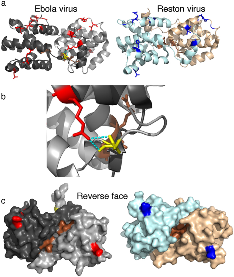
Figure 3. SDPs present in the VP30 dimer.
The dimer structure of both Ebola virus (PDB structure 2I8B) and Reston virus (PDB structure 3V7O) VP30 are shown with SDPs indicated (red – Ebola virus, blue – Reston virus) and functional residues (brown – A179, K180). (a) Cartoon representation: For the Ebola virus the hydrogen bond of R262 with the residue 141 of the other subunit is shown. (b) enlarged display of the hydrogen bond between R262 and the backbone of residue 141. (c) Surface representation of the reverse face of the dimer from A, showing the location of the functional residues A179 and K180 within the dimer

