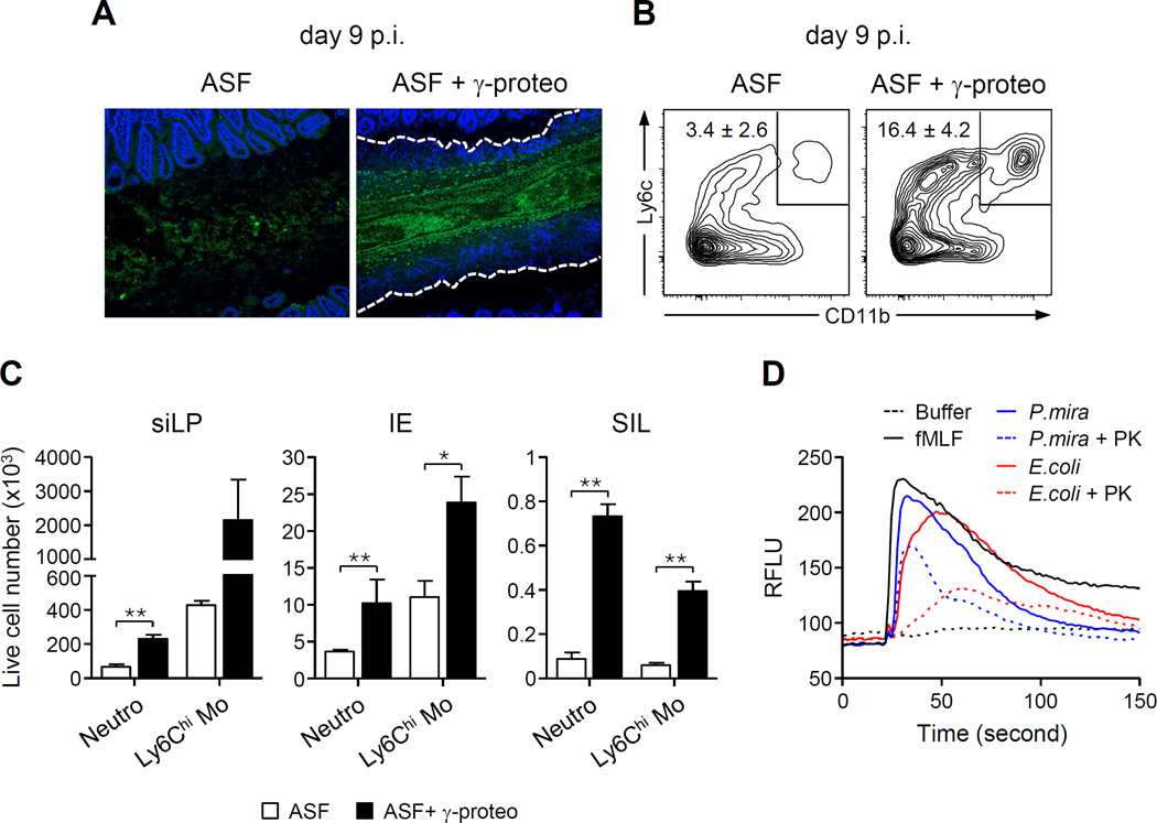
Figure 5. γ-Proteobacteria promote luminal recruitment of inflammatory cells.
Germ-free mice were colonized with either ASF bacteria or ASF + γ-proteobacteria and then infected with T. gondii. (A) Representative image of 16S FISH staining of ileal sections. The white dashed lines represent the border of the cellular casts. (B) Contour plots show the percentage of CD11b+Ly6C+ leukocytes isolated from the lumen. Numbers indicate the mean ± SEM percent inside the gate. (C) Number (mean ± SEM) of inflammatory monocytes and neutrophils isolated from the small intestine lamina propria, IE and lumen (SIL) of ASF or ASF + γ-proteobacteria mice. (D) Supernatants isolated from E. coli and P. mirabilis (P.mira) cultures were tested for their ability to induce calcium flux in neutrophils. Histogram indicates the calcium flux over time from purified neutrophils, treated as indicated (fMLF = N-formyl methionine leucyl phenylalanine; PK = proteinase K). See also Figure S4.

