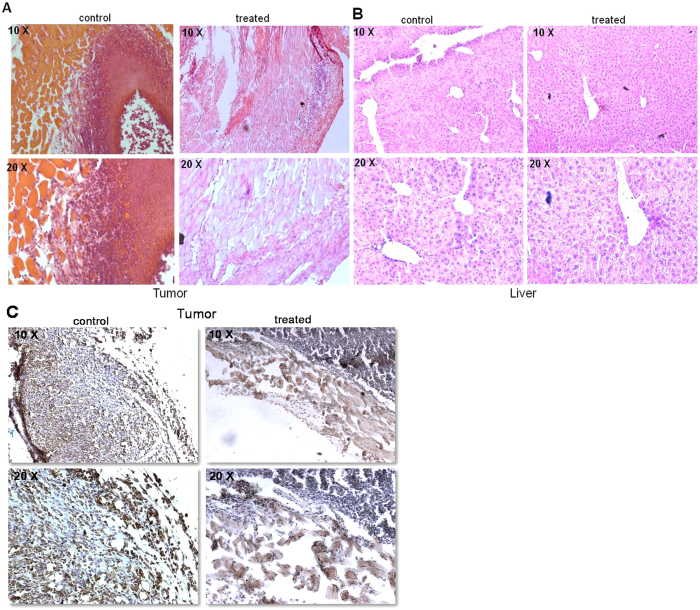
Figure 7. Histological and immunohistochemical analysis of VCE treated tumor mice.
(A,B) Haematoxylin and Eosin (HE) stained sections prepared from either tumor tissues (A) or liver (B), which were from VCE treated mice for 25 days or untreated tumor control animals. Images represent HE stained sections of control tumor and VCE treated tumor (A); control liver and VCE treated liver (B). (C) Antibody staining for Ki67 on 25th day tumor tissue and VCE treated tumor tissue. Images are shown in both 10× and 20× magnification.

