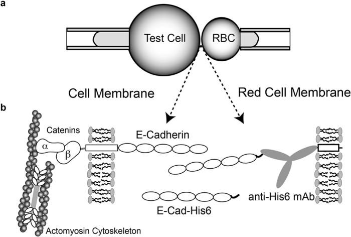
Figure 2. Illustration of the Micropipette Setup.

a) A test cell (for example, A431D cells) expressing full-length cadherin is aspirated into the left micropipette, and a Red Blood Cell (RBC) ectopically modified with E-Cad-His6 is aspirated into the right micropipette b) Protein orientations on the opposing test cell and the RBC. The RBC is covalently modified with anti-His6 antibodies, which capture and orient the histidine tagged E-Cadherin extracelluar domains. The cells are repetitively brought into contact for a defined period (and contact area) and retracted with piezoelectric manipulators. Adhesion events are quantified from visible RBC deformations and recoil upon bond failure. In the left micropipette, cells can be replaced with a modified RBC, as in the right pipette, in order to quantify binding between ectodomains only.
