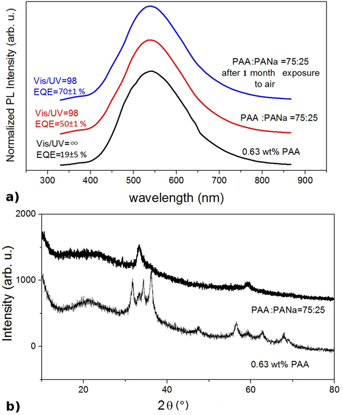
Figure 10.

(a) PL spectra of ZnO nanoparticles synthesized by the hydrolysis of ZnEt2 with 0.63 wt% of PAAH only (bottom curve) and with PAAH and PAANa of 3:1 (middle curve) and after 2 months exposure to air (top curve). (b) XRD diffractograms of ZnO nanoparticles synthesized by the hydrolysis of ZnEt2 with 0.63 wt% of PAAH only (bottom curve) and with PAAH and PAANa of 3:1 (top curve). The plots are shifted along the y-axis for clarity. (c) HRTEM of small ZnO nanocrystals. The wurtzite structure is clearly seen from the inset showing the FFT of the image.
