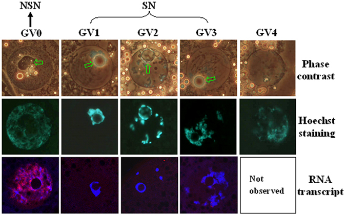
Figure 1. Photographs of porcine oocytes showing different germinal vesicle (GV) chromatin configurations and global RNA transcription.

Photographs in the top and middle rows for each chromatin configuration are the same oocyte observed with phase contrast and fluorescence, respectively, after Hoechst 33342 staining. The nucleolus is indicated with arrows in the phase contrast images. Original magnification ×400. For convenience, GV-0 was designated as NSN configuration, and GV1, GV2 and GV3 were classed as SN configuration in the present study. Photographs in the bottom row are laser confocal (merged) images showing global RNA transcription of porcine oocytes with different GV chromatin configurations. DNA and RNA were pseudo colored blue and red, respectively. Original magnification ×630. Each treatment was repeated 3 times with each replicate containing about 30 oocytes.
