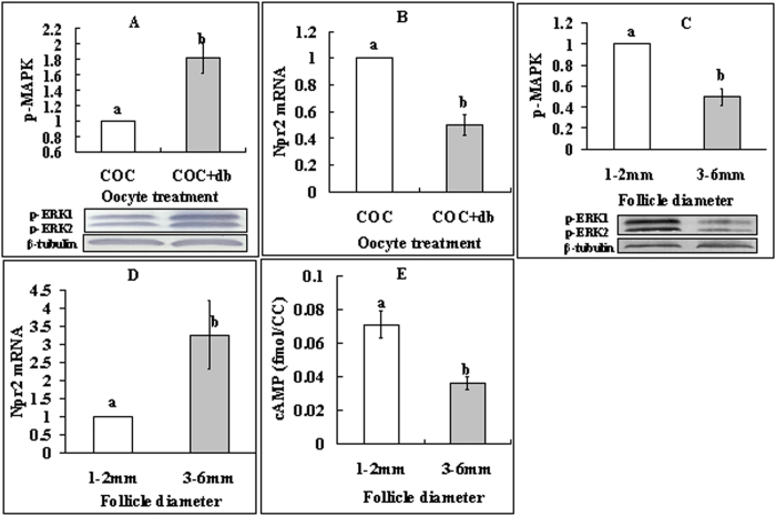
Figure 4. Levels of p-MAPK, Npr2 mRNA and cAMP in CCs.
(A,B) show p-MAPK and Npr2 mRNA, respectively, in CCs from COCs that had been cultured for 16 h with (+db) or without db-cAMP. (C–E) show p-MAPK, Npr2 mRNA and cAMP, respectively, in CCs from follicles of different diameters. Each treatment was repeated 3 times with each replicate containing CCs from 200 COCs for p-MAPK and Npr2 mRNA assays and about 2 × 10⁵ CCs for cAMP measurement. (a,b) Values without a common letter above their bars differ significantly (P < 0.05).

