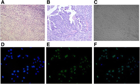
Fig. 2.

Culturing of primary human ovarian cancer cells. a Pathological section of normal human ovarian tissue with benign pathology (H&E stained, ×100). b Pathological section of human ovarian cancer tissue (H&E stained, ×100) that was classified as serous ovarian adenocarcinoma (I-II grade) according to WHO criteria. c Representative morphology of ovarian carcinoma cells grown in primary culture medium (×100). d Cultured primary human ovarian cancer cells stained with nuclear dyes-Hochest33258 (×100). e Cultured primary human ovarian cancer cells stained with anti-cytokeratin 7-FITC. f The confocal of D and E pictures
