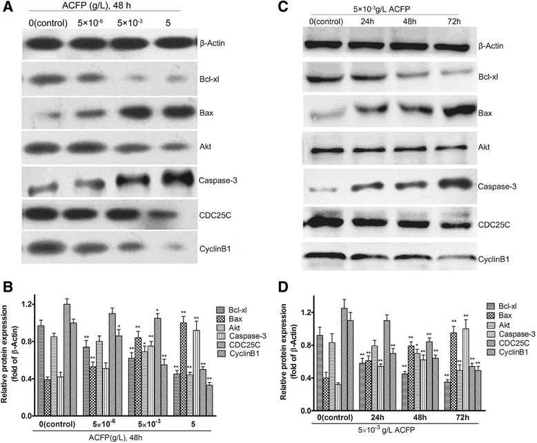
Fig. 7.

ACFP promotes changes in Bcl-xl, Bax, Akt, Caspase-3, CDC25C and CyclinB1 protein levels in primary human ovarian cancer cells. a Western blot analysis was performed after 48 h ACFP treatment in primary human ovarian cancer cells and antibodies specific to Bcl-xl, Bax, Akt, Caspase-3, CDC25C and CyclinB1 were used to assess protein levels. The β-Actin was used to show the similar amount of protein loaded in different lanes. b The relative intensities of protein bands in A were determined using Quantity-One software version 4.62 (Bio-Rad, USA) and normalized using β-Actin band intensity. c After 5 × 10⁻³ g/L ACFP treatment for different time, Bcl-xl, Bax, Akt, Caspase-3, CDC25C and CyclinB1 were detected. d The relative intensities of protein bands in c were determined using Quantity-One software and normalized using β-Actin band intensity. The data in b and d are shown as means ± SD of 12 primary ovarian cancer cells measurements from 12 patients, * P < 0.05, ** P < 0.01 compared with control (the vehicle group)
