Fig. 8.

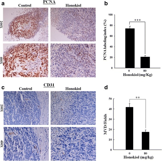
Honokiol markedly decreases the immunohistochemical staining of PCNA and CD31 in SAS SP xenograft tumor tissue. Immunohistochemistry staining of PCNA and CD31 was performed in the paraffin-embedded tissue sections of tumors from mice treated with or without honokiol (80 mg/kg). a The staining intensity (brown color) of PCNA was markedly lower in honokiol-treated group. b The PCNA labeling indexes of control and honokiol-treated groups. c The staining intensity of CD31 (endothelial cell marker) was markedly lower in honokiol-treated group. d The number of CD31 positive microvessel was counted at 200x magnification under a microscope. Significantly reduced microvessel density (MVD)/fields was observed in honokiol-treated group. Data shown are mean ± SD. **p < 0.01; ***p < 0.001, significant difference vs. control
