
Fig. 4.
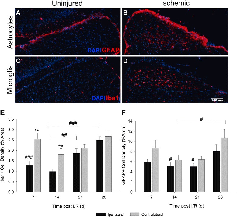
Retinal I/R activates and increases glial cell populations in the SC. Representative images demonstrate differences observed in astrocytes (a), (b) and microglia (c), (d) between uninjured ipsilateral and I/R-receiving contralateral hemispheres of the SC. (e) Quantification of microglial density was observed to be significantly different between hemispheres on days 7 and 14 (p < 0.01) following I/R. No differences were detected between hemispheres by 21 days post injury. However, a statistically significant difference was determined in the uninjured ipsilateral hemisphere when days 21 (p < 0.01) and 28 (p < 0.001) were compared to earlier time points. (f) Density of GFAP+ astrocytes was quantified; however, no significant differences could be determined between SC hemispheres at any time point following retinal I/R. However, statistical differences (p < 0.05) were observed in both ipsilateral and contralateral hemispheres when day 28 was compared against astrocyte density on days 14 and 21. Mean ± SEM, n = 7 per group. **p < 0.01 using students paired t-test between ipsilateral and contralateral hemispheres, #p < 0.05 ##p < 0.01 ###p < 0.001 across time points determined by One-way ANOVA followed by Holm-Sidak post test. Scale bars = 100μm