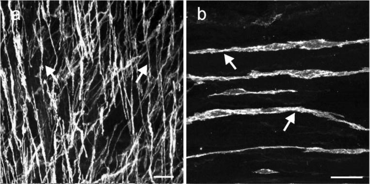
Fig. 1.

ICC in the lower esophageal sphincter. (a, b) digital reconstructions of confocal images from flat cryosections (100 μm) of the LES. Kit+ ICC (arrows) possessed spindle shaped morphology with a central oval nucleus and were interspersed within the circular and longitudinal muscle layers. The dense population of ICC-IM ran parallel to the longitudinal axis of the muscle fibers. Scale bars = 50 μm in (a) and 25 μm in (b)
