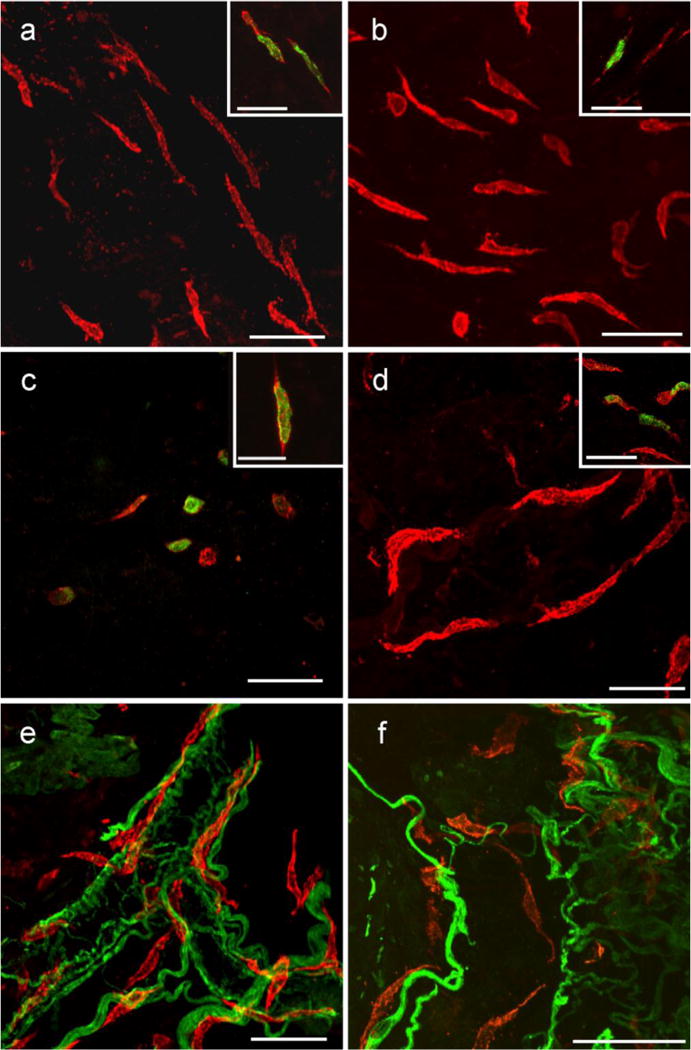
Fig. 6.

Kit+ cells within the submucosa and lamina propria of the GI tract. Flat sections and whole mount preparations of the mucosa and underlying submucosa from stomach, small intestine and colon. Kit+ cells within the submucosa and lamina propria in the gastric fundus (a), antrum (b), small intestine (c) and colon (d) were spindle shaped and did not appear to have a distinct orientation except when in close association with submucosal blood vessels (e,f colon shown). In the small intestine the number of these Kit+ cells that were spindle shaped was less than in other organs, but a larger number of rounded Kit+ cells were observed in the intestinal submucosa. Double labeling with PGP 9.5 and Kit revealed that the spindle shaped cells ran close to but were not intimately associated with nerves. PGP 9.5 labeling also identified autonomic nerves associated with submucosal blood vessels and spindle shaped cells were closely associated with these vessels (e). Double labeling with Kit and histamine revealed a sub-population of these spindle shaped cells were histamine+ in the stomach, small intestine and colon, suggesting these cells were likely mast cells (insets in a–d). Scale bar in (o) = 50 μm and applies to all panels. Scale bar in the inset in (i) = 50 μm and applies to insets in (g–i). Scale bar in all panels and insets = 50 μm, except inset in (c) = 25 μm
