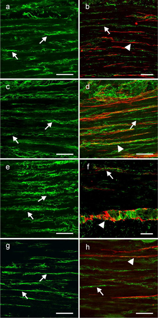
Fig. 7.

PDGFRα is expressed in a separate population of interstitial cells that are not Kit+. (a,c,e,g) Shows the cellular distribution of PDGFRα within the circular muscle layer of the gastric fundus, antrum small intestine and colon, respectively. PDGFRα + cells (arrows) ran parallel to the long axis of the circular muscle fibers. (b,d,f h) show double labeling of PDGFRα (green; arrows) and Kit (red; arrow heads) in two populations of cells in the fundus, antrum, small intestine and colon, respectively. Although the two cell populations were closely apposed to one another they were distinct, providing evidence that PDGFRα+ cells were not Kit+ ICC. Scale bars in all panels = 50 μm
