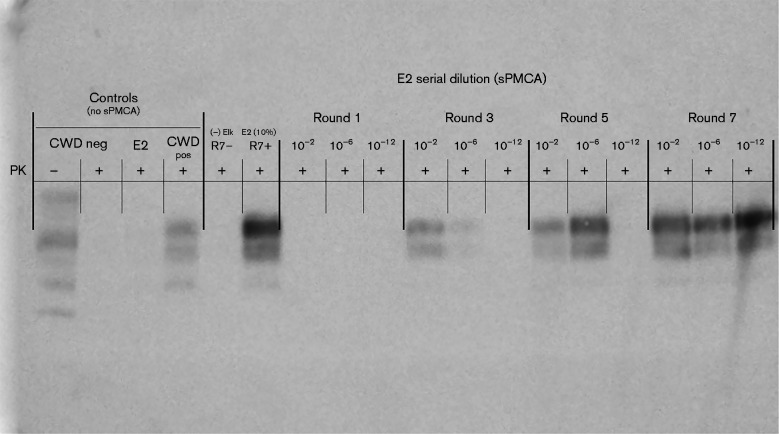
Fig. 1.
PrPCWD detection in dilutions of CWD-positive elk brain following sPMCA. Representative Western blots for detection of PrPCWD in CWD-positive elk brain homogenate (E2) following sPMCA (dilutions 10− 2, 10− 6 and 10− 12, rounds 1, 3, 5 and 7 shown). PrPCWD was not detected in a 10 % homogenate of E2 prior to sPMCA or after one round of sPMCA. After three and five rounds of E2 sPMCA, PrPCWD was detected at 10− 6. PrPCWD was detected at 10− 12 following seven rounds of sPMCA. sPMCA controls (R7– and R7+) showed complete proteinase K (PK) digestion of negative elk brain homogenate (R7–) and proteinase K-resistant PrPCWD in E2 CWD-positive elk brain homogenate (R7+) (10 % homogenates, undiluted, round 7). Western blot controls (no sPMCA) showed complete proteinase K digestion of PrP in negative white-tailed deer brain homogenate and proteinase K-resistant PrPCWD in CWD-positive white-tailed deer brain homogenate (10 % homogenates, undiluted, no sPMCA).

