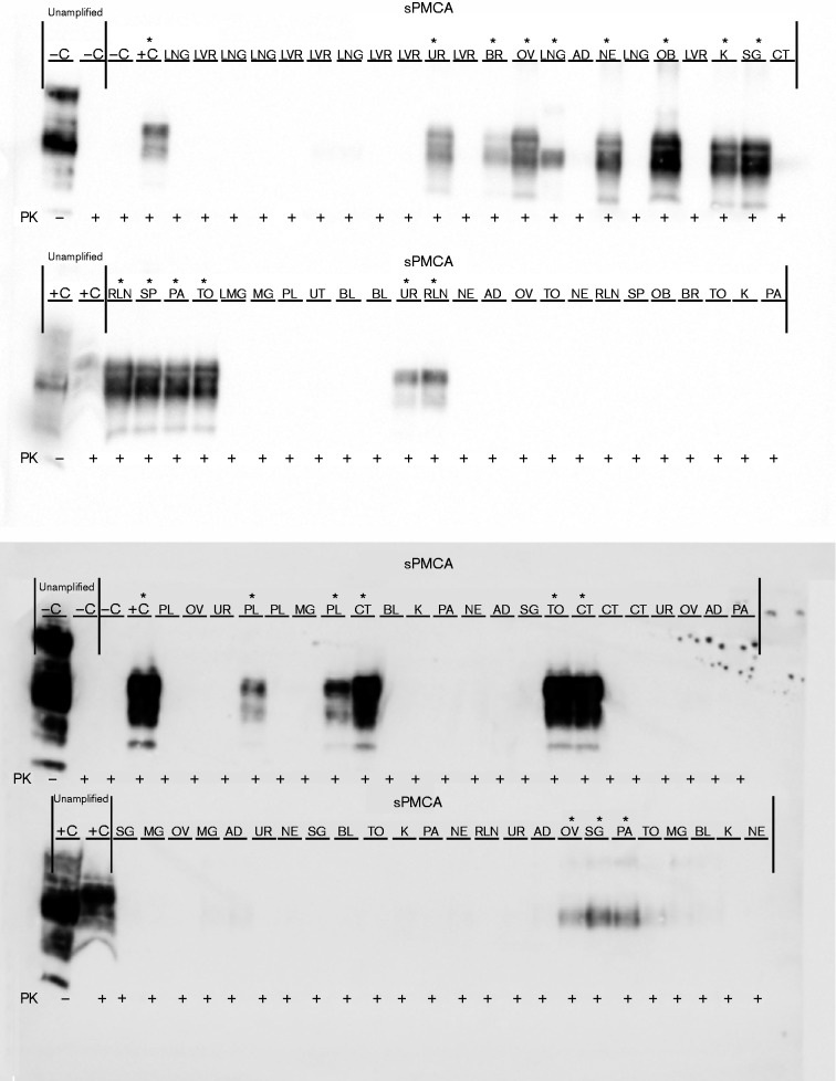
Fig. 5.
PrPCWD detection in elk dam tissues following seven rounds of sPMCA. Representative Western blots for detection of PrPCWD in dam tissues. sPMCA controls showed the lack of amplification for a CWD-negative sample and the successful amplification of a known CWD-positive white-tailed deer. Western blot controls (no sPMCA) showed complete proteinase K (PK) digestion of PrP in a negative white-tailed deer brain homogenate and PrPCWD signal post-proteinase K digestion in a CWD-positive white-tailed deer brain homogenate (10 % homogenate, undiluted, no sPMCA). Asterisks denote sPMCA + CWD amplification. Tissue codes: C, control. (+, positive; –, negative); AD, adipose tissue; BL, urinary bladder; BR, brain; CT, cotyledon; K, kidney; LNG, lung; LVR, liver; MG, mammary gland; NE, nasal epithelium; OB, obex; OV, ovary; PA, pancreas; PL, placenta; RLN, retropharyngeal lymph node; SG, salivary gland; SP, spleen; TO, tongue; UR, ureter; UT, uterus.

