Figure 2.

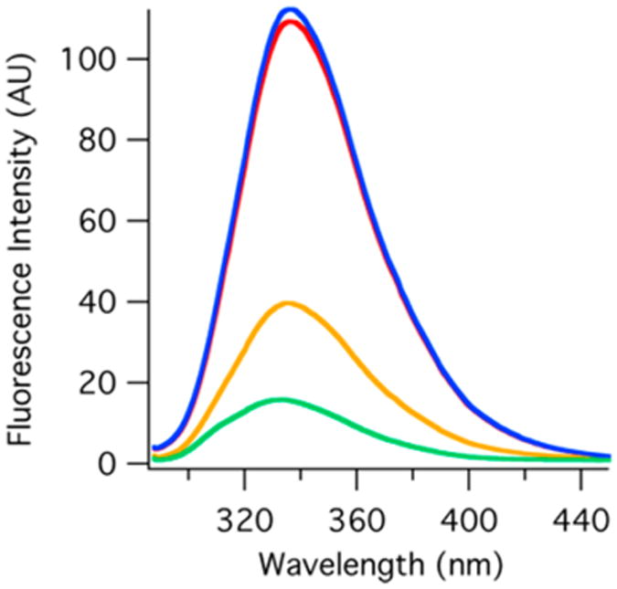
Relative equilibrium tryptophan fluorescence (excitation at 280 nm) of the DHFR apoenzyme (red), DHFR·folate (yellow), DHFR·NADP+ (blue), and DHFR·NADP+·folate (green). These spectra were recorded at 20 °C with 3 μM enzyme.

Relative equilibrium tryptophan fluorescence (excitation at 280 nm) of the DHFR apoenzyme (red), DHFR·folate (yellow), DHFR·NADP+ (blue), and DHFR·NADP+·folate (green). These spectra were recorded at 20 °C with 3 μM enzyme.