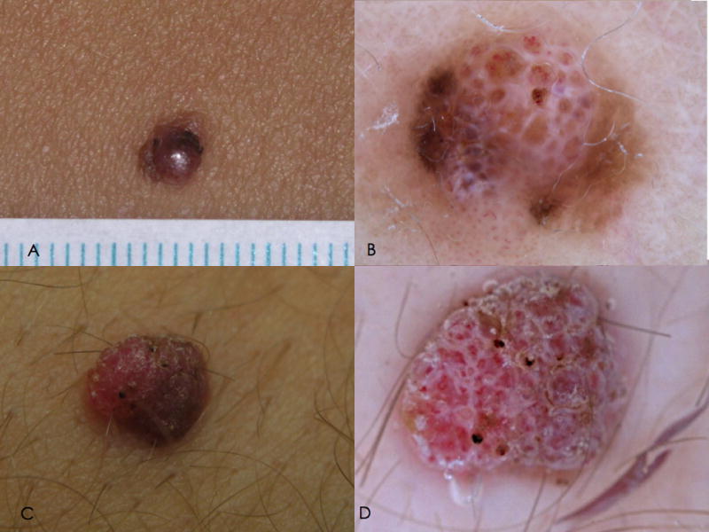
Figure 2.

Clinical and dermoscopic features of hypomelanotic AST (atypical Spitz tumor). A. Clinical appearance of a hypomelanotic nodule arising on the lower limb of a 34 year-old woman. B. On dermoscopy the lesion exhibits a multicomponent pattern, with homogeneous brown pigmentation, white lines and polymorphic vessels.
C. Clinical appearance of a hypopigmented nodule arising on the leg of a 22 year-old boy. D. On dermoscopy, nonspecific pattern is detected, with polymorphic vessels, brownish pigmentation, and ulceration.
