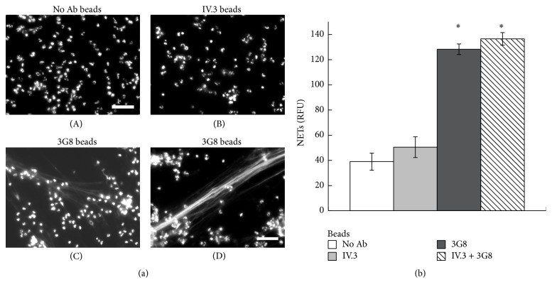
Figure 11.
Anti-FcγRIIIb-opsonized particles induce NET formation. (a) Human neutrophils (PMN) mixed with latex particles nonopsonized (no Ab beads) or opsonized with monoclonal antibody IV.3, anti-FcγRIIa (IV.3 beads), or with monoclonal antibody 3G8, anti-FcγRIIIb (3G8 beads), were incubated for four hours and fixed and stained for DNA (DAPI). Microphotographs were taken at 200x magnification and are representative of three experiments. Bar is 50 μm. (b) The relative amount of NETs was estimated from SYTOX Green fluorescence in relative fluorescent units (RFU) at 4 hours after stimulation, as described in Materials and Methods. Data are mean ± SEM of four experiments. Asterisks denote conditions that are statistically different from control (p < 0.02).

