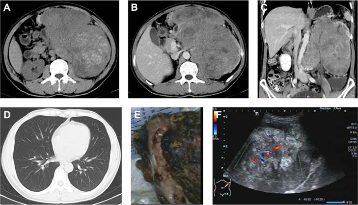
Figure 2.
A 43-year-old male with abdominal mass.
Notes: (A) An unenhanced CT image shows a large mass in the left kidney with hemorrhage. (B) A contrast-enhanced CT image shows a thrombus in the left renal vein. (C) A sagittal-reformatted image shows the enlargement of the aortocaval lymph node compressing the artery. (D) A lung window setting shows the presence of metastatic lung nodules. (E) A photograph of a gross specimen shows a white and gray mass with hemorrhage and necrosis. (F) A Doppler image shows an ill-defined heterogenous hyperechoic mass with a little vascularity in the left kidney.
Abbreviation: CT, computed tomography.

