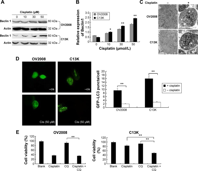
Figure 2.
Cisplatin induces cytoprotective autophagy in cisplatin-resistant ovarian cancer cells.
Notes: Cells were treated with different concentrations (0–50 μmol/L) of cisplatin for 48 hours. (A) Cell lysates were collected to detect the protein levels of Beclin1. (B) The messenger RNA level of Beclin1 was determined using a real-time polymerase chain-reaction kit. Data represent the mean and standard deviation values from three independent experiments (*P<0.05, **P<0.01). (C) Cells were treated with 50 μmol/L cisplatin for 48 hours and subjected to electron-transmission microscopy for the detection of autophagic vacuoles; the black arrows indicate the autophagic vacuoles (magnification 10,000×, scale bar 2 μm). (D) Analysis of LC3 aggregation in ovarian cancer cells by fluorescence microscopy. Cells transiently transfected with GFP–LC3 using Lipofectamine 2000 were cultured in Roswell Park Memorial Institute medium with 10% fetal bovine serum in the absence or presence of 50 μmol/L cisplatin for 48 hours. Accumulation of GFP–LC3 puncta was observed (scale bar 40 μm). Quantitation of the GFP–LC3 puncta was performed by counting 20 cells for each sample, and the average number of puncta per cell are shown. Bars represent the mean ± standard deviation values from three independent experiments (**P<0.01). (E) Cells were pretreated with chloroquine (CQ; 50 μmol/L) for 24 hours, and then treated with cisplatin (50 μmol/L) for 48 hours. Cell viability was determined using the CCK-8 assay (**P<0.01).

