Figure 4.
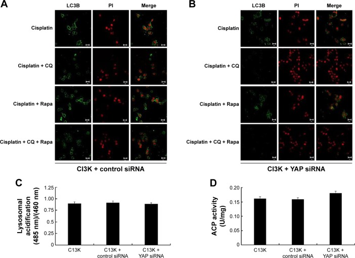
YAP interferes with autophagic flux by enhancing autolysosome degradation in cisplatin-resistant ovarian cancer cells.
Notes: Without treatment, control small interfering RNA (siRNA) and YAP siRNA C13K cells were cultured for 48 hours. An immunofluorescence assay was performed to monitor autophagy-related morphological changes. C13K plus control siRNA (A) or YAP siRNA (B) cells were subjected to different treatments: cisplatin, chloroquine (CQ) + cisplatin, rapamycin (Rapa) + cisplatin, and CQ + Rapa + cisplatin. The presence of autophagosomes was detected by using the LC3B antibody and fluorescence microscopy. Nuclei were counterstained with propidium iodide (LC3B green, PI red); scale bar 20 μm. (C) Comparison of lysosomal acidification in cells based on the ratio of the emission intensity at 520 nm and at the two excitation wavelengths 485 nm and 460 nm. Results expressed as mean ± standard error values from three independent experiments. (D) Acid phosphatase (ACP) activity of the C13K cells was assayed. Data shown as mean ± standard error.

