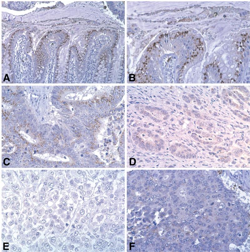
Fig. 1.
CaSR expression in normal colonic epithelium and in colon carcinoma. A and B. In the normal crypt, there is intense staining of the differentiated cells in the upper part of the crypt. C. In an area of moderately-differentiated tumor, weak and variable staining is observed. D. There is essentially no staining of isolated cells at the invasive front. Likewise, there is little or no staining of invasive cells even where there is rudimentary glandular structure. E and F. In areas of histologically undifferentiated tumor (no glandular structure evident; only sheets of undifferentiated tumor cells), there is no CaSR staining (See Chakrabarty et al. 2003; 2005 for details).

