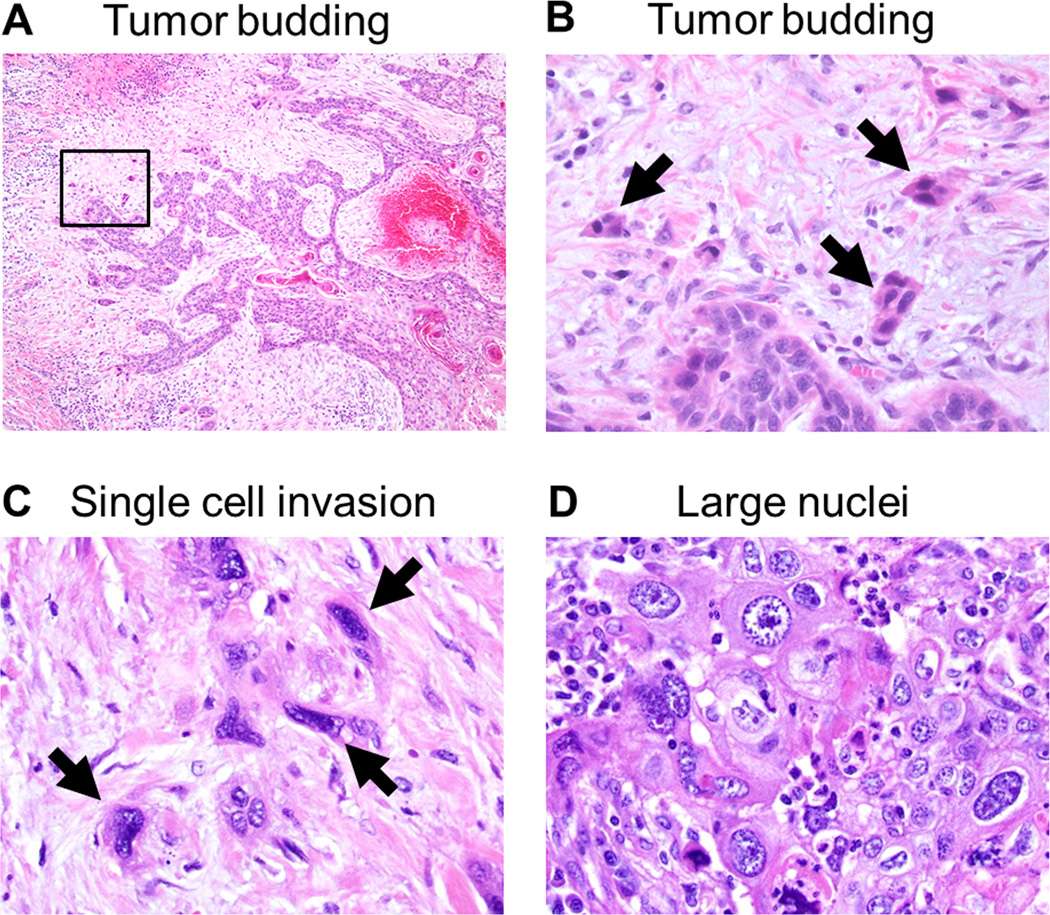
FIGURE 2.
Tumor budding and single cell invasion (hematoxylin and eosin-stain; original magnification, ×40: A, ×400: C–D).
(A) Tumor budding identified in invasive tumor edge. (B) Higher magnification of a square box in the Figure 3A showing tumor budding composed of less than 5 tumor cells (arrows). (C) Single cell invasion of tumor cells in stroma (arrows). (D) Large nuclei defined as >4 small lymphocytes in diameter.

