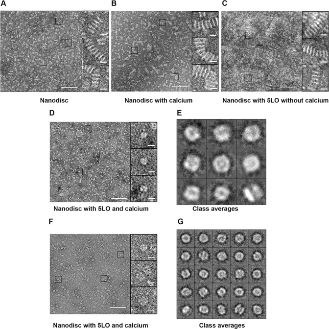
Fig 4. Both indirect and direct visualization of the effect of calcium on interaction of monomeric 5LO with ND, by negative stain electron microscopy.
(A), ND showing stack formation induced by PTA stain, homogeneous size and the rigid structure of ND. (B), the ND preparation as in A but in the presence of Ca2+ shows the same amount of stacking as in A. (C), PTA-stained samples of equimolar concentrations of ND incubated with 5LO in calcium-free buffer, clearly showing stack formation, which signifies lack of interaction between 5LO and ND. (D), PTA stained samples of equimolar concentrations (0.8 μM) of ND incubated with 5LO in the presence of 1 mM Ca2+, showing very few stacks. A lack of PTA-induced stacking formation, signifies the interaction between 5LO and ND. The magnifications on the right in are representatives of the majority of the particles found in the corresponding image. (E), Class averages of 5LO-ND-complexes stained with PTA. Five images were used (including the one in D) to box 377 particles. Box-size is 20.8 nm. Scale bars are 10 nm and 100 nm respectively. (F), PTA stained samples of ND (0.8 μM) incubated with 5LO (1.6 μM) in the presence of 1 mM Ca2+, showing even fewer stacks. Most particles are larger and have different shapes compared to the 1:1 ratio shown in (D). (G), Class averages of 5LO-ND-complexes stained with PTA. Thirty six images were used to box 573 particles. Box-size is 26.6 nm. Scale bars are 10 nm and 100 nm respectively.

