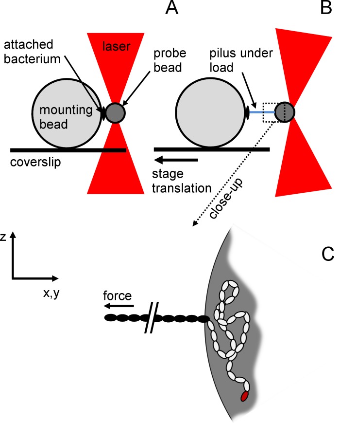
Fig 2. Experimental configuration of single-molecule force spectroscopy assays.
Scheme illustrating the measurement configuration that was used during the experiments with no force applied (A) and under force (B) when the stage was moving. C is a close-up of B.). The pilus attached to the trapped bead is disproportionally long for reasons of depiction. The 10.5-μm mounting bead (MB) was immobilized on the coverslip while the 1-μm probe bead (PB) was trapped by optical tweezers (OT). A piliated bacterium was non-specifically attached to the MB and a pilus to the PB. When the coverslip was moved, and the trap kept in a fixed position, a force was directly exerted on the pilus. (C) Assuming adhesion to be non-specific, the most likely situation is that a portion of the pilus was attached (white subunits) and not solely the adhesion pilin (red subunit). Only a part of the pilus (black subunits) was thus subjected to the applied force.

