Figure.

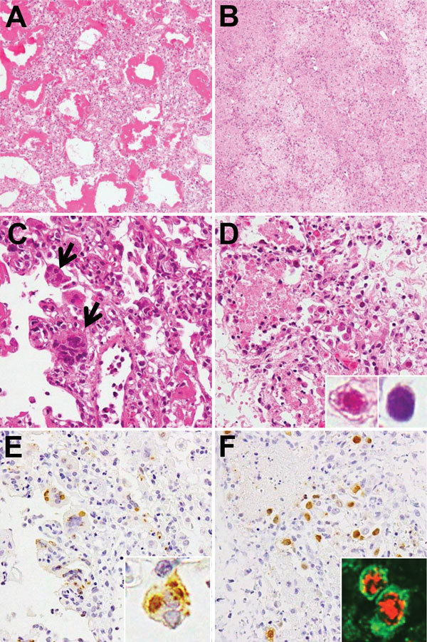
Histologic findings from postmortem lung tissues of children who died from measles-associated pneumonia in a pediatric intensive care unit, National Hospital of Pediatrics, Hanoi, Vietnam, January–October 2014. A) Diffuse alveolar damage with hyaline membrane formation (hematoxylin and eosin [H&E] stain, original magnification ×100). B) Necrotizing pneumonia with coagulation necrosis (H&E stain, original magnification ×100). C) Measles giant cell pneumonia. Arrows indicate syncytial cells with intracytoplasmic and intranuclear eosinophilic inclusions that were observed in the thickened alveolar walls (H&E stain, original magnification ×400). D) Adenovirus (AdV) pneumonia with necrotic epithelial cells and intranuclear inclusion bodies. Inset shows eosinophilic inclusion with halo and basophilic inclusion without halo (H&E stain, original magnification ×400). E) Measles nucleoprotein (brown) detected by immunohistochemistical analysis. Inset shows inclusions in syncytial cells with measles nucleoprotein (original magnification ×400). F) AdV antigen (brown) detected by immunohistochemistry (H&E stain, original magnification ×400). Inset: AdV antigens (red) were detected in the epithelial membrane antigen (green)–positive pneumocytes (double immunofluorescence stain, original magnification ×400).
