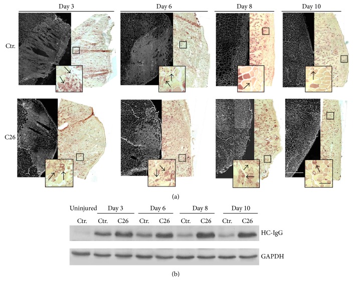
Figure 2.
C26-bearing mice show a prolonged inflammation response during muscle regeneration. (a) Left panels: immunofluorescence analysis of IgG expression performed at the lesion site of reconstructed areas of tibialis muscles from both control and C26-bearing mice. (a) Right panels: esterase staining uptake at the same lesion sites highlighting the presence of macrophages in the inflamed muscles. Insects depict magnification of areas defined in the squares. Black arrows show macrophages. White bar = 0.5 mm; black bar = 100 μm. (b) WB analysis of IgG expression on extracts from muscles shown in (a). First lane was loaded with the extract from a healthy, uninjured control muscle. GAPDH was used as loading control.

