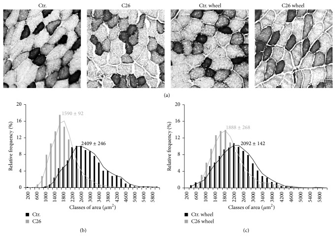
Figure 6.
Voluntary wheel running rescues skeletal muscle atrophy in C26-bearing mice. (a) NADH staining in TA muscles. Glycolytic fibers are shown as pale colored while oxidative fibers stain as dark. Bar = 100 μm. Morphometric analysis of glycolytic fibers among healthy controls (black bars) and C26-bearing mice (gray bars) at rest (b) and in the presence of voluntary wheel running (c). Numbers represent the median value ± SEM of three independent experiments (C26 versus C26 wheel: F = 1126; p = 0.0001).

