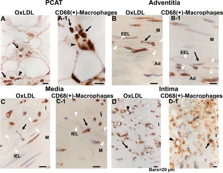
Fig 2. Comparison of dotted oxLDL deposits and CD68 (+)-macrophages.
Adjacent sections were stained immunohistochemically for oxLDL or CD68(+)-macrophages for comparison. Both oxLDL deposits and macrophages showed round (arrowhead in A, A-1) or spindle-like configuration (arrows in A, A-1; B, B-1; C, C-1; D, D-1). Dotted deposits of oxLDL and macrophages (arrows in B, B-1, C, C-1) traversing external elastic lamina (EEL; white arrowheads in B, B-1) and internal elastic lamia (IEL; white arrowheads in C, C-1) were demonstrated, strongly suggesting that oxLDL was carried by CD68(+)-macrophages and migrated from adventitia to media, and then to intima. Vacuole-like structures surrounded by oxLDL or CD68 were also observed in intima (arrowheads in D, D-1), suggesting that they are foam cells. Scale9 bars = 20 μm.

