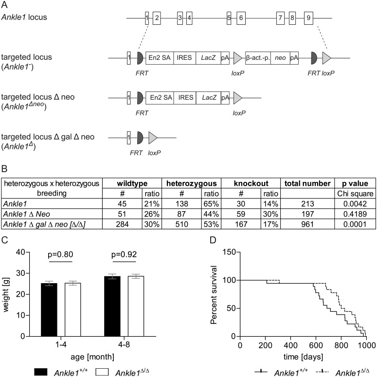
Fig 2. Ankle1 knock-out mouse model.
(A) Schematic drawing of the Ankle1 wild-type locus with exons 1 to 9, the knock-out allele carrying the complete deletion cassette (Ankle1-) consisting of FRT recombination sites (semi circle), splice acceptors site (En2 SA), Internal Ribosome Entry Site (IRES), LacZ gene (LacZ), polyadenylation site (pA), loxP recombination site (triangle), β-actin promoter (β -act.-p.) and aminoglycoside phosphotransferase gene (neo) and the knock-out allele after CRE-recombination (Ankle1Δneo) carrying the reporter gene cassette only and the knock-out allele after FLP-mediated recombination (Ankle1Δ); (B) Birth statistics of different Ankle1-deficient mouse strains (for organization of the genomic locus see Fig 2A) from heterozygous pairings, for determination of statistical significant differences between the observed and the expected birth ratios (25% wildtype, 50% heterozygous, 25% knock-out) a Chi square test was performed, p≤0.05 statistical significant, p≤0.01 very statistical significant; (C) Weight of Ankle1+/+ and Ankle1Δ/Δ animals 1–4 months and 4–8 months old, paired students t-test, statistical significance p≤0.05, 17 ≤ n ≤ 19; (D) Survival curve for Ankle1+/+ and Ankle1Δ/Δ mice, log rank (Mantel-Cox) test p = 0.11, n = 18.

