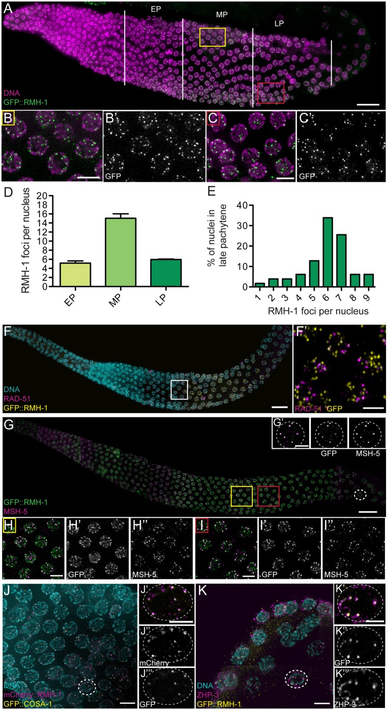
Fig 3. Dynamic localization of RMH-1 to distinct foci in pachytene.
(A) In early pachytene (EP), GFP::RMH-1 is diffuse and few foci are detected on DNA. In mid pachytene (MP), high numbers of foci (up to 25 per nucleus) are observed (yellow square and B and B′). In late pachytene (LP), GFP::RMH-1 concentrates into bright foci, on average six per nucleus (red square and C and C′). (D) Quantification of the average number of RMH-1 foci per nucleus in EP, MP, and LP (n = 123 nuclei for EP, n = 205 for MP, and n = 180 for LP). Data are represented as mean +/- SEM. (E) Histogram showing the percentage of late pachytene nuclei containing a certain number of RMH-1 foci per nucleus (one to nine) (n = 180 nuclei). (F) Staining for RAD-51 and GFP::RMH-1. RMH-1 localization starts after and persists longer than the RAD-51 positive zone. (F′) GFP::RMH-1 and RAD-51 mark different recombination intermediates, as they do not colocalize. (G–I) Staining for MSH-5 and GFP::RMH-1. (H–H′′) RMH-1 and MSH-5 partially colocalize in MP (yellow square). (I–I′′) Both proteins become enriched at brighter foci as pachytene progresses (red square). (J) Staining for COSA-1::GFP and mCherry::RMH-1. (K) Staining for ZHP-3 and GFP::RMH-1. In LP, RMH-1 colocalizes at CO sites with MSH-5 (G′), COSA-1 (J′–J′′′) and ZHP-3 (K′–K′′′). Scale bar = 20 μm for gonads, 5 μm for pachytene nuclei insets, and 2.5 μm for single nucleus.

