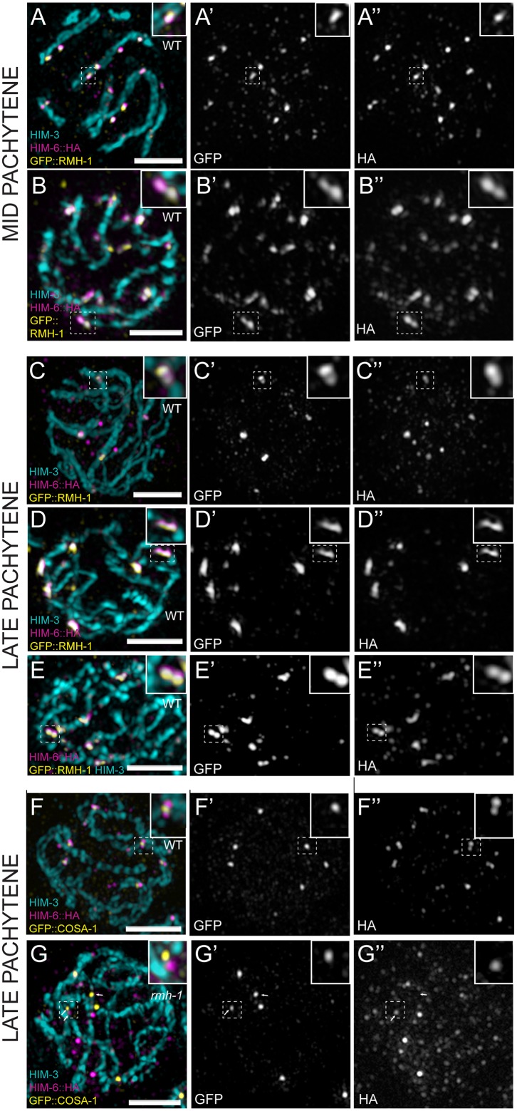
Fig 6. Foci of RMH-1 and BLM are resolved as doublets or elongated structures during the pachytene stage.
(A–G) Single pachytene nuclei imaged by SIM. (A–B) In the WT, HIM-6 and RMH-1 colocalize in MP. Foci appear elongated or as a doublet (see insets). (C–E) In LP, RMH-1 and HIM-6 are concentrated at CO sites contained in a structure resolvable into a doublet (see insets). (F) Colocalization of HIM-6 and COSA-1 at CO sites in WT. (G) In rmh-1(jf54), HIM-6 does not colocalize with COSA-1 at CO sites (white arrow) but can be found in close proximity (white arrows). Scale bar 2μm.

