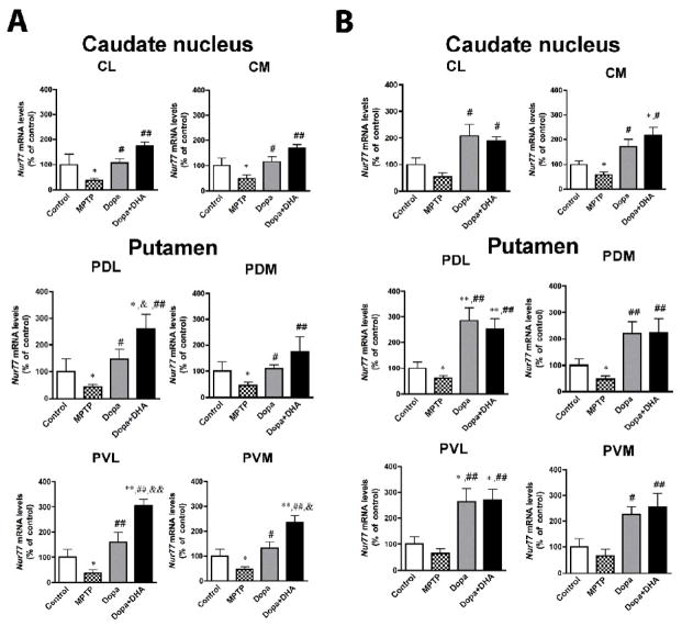
Figure 2.
Modulation of Nur77 mRNA levels in L-Dopa-treated MPTP monkeys in the caudate-putamen. Nur77 mRNA levels were measured in control, MPTP, MPTP + L-Dopa and MPTP + L-Dopa + DHA treated monkeys in the anterior (A) and posterior (B) caudate nucleus and putamen. Nur77 mRNA levels were evaluated in lateral (CL) and medial (CM) portions of the caudate nucleus as well as in dorsolateral (PDL), dorsomedial (PDM), ventrolateral (PVL) and ventromedial (PVM) portions of the putamen. Values are expressed in percent (%) of control values. Absolute values in nCi/g tissue of controls are presented in Table 2. Histogram bars represent means ± SEM (N=4–5 per group) (* p < 0.05 and ** p < 0.01 vs Control, # p < 0.05 and ## p < 0.01 vs MPTP and & p < 0.05 and && p < 0.01 vs MPTP + L-Dopa).

