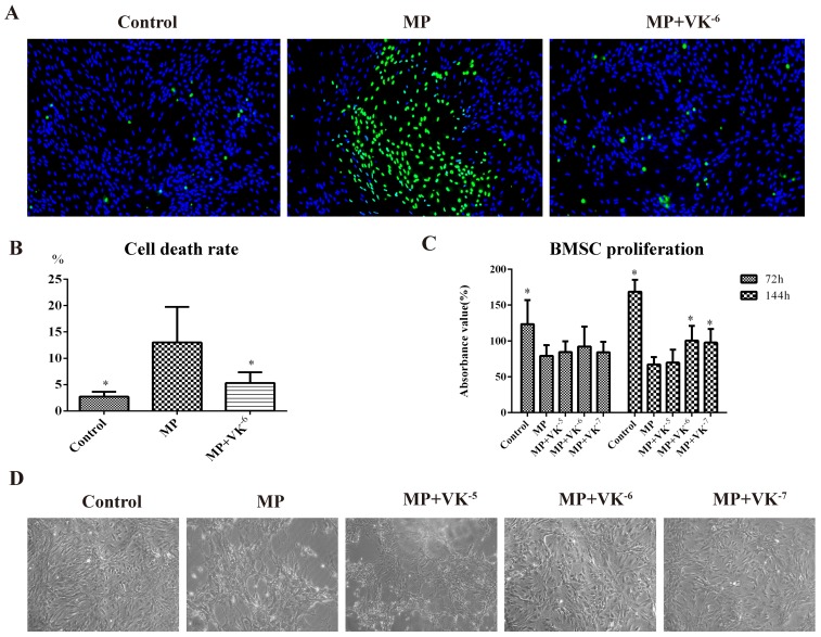
Figure 1.
Effects of MP and MP supplemented with VK2 on the proliferation and survival of BMSCs. (A, B) Cell viability staining was performed 144 h after incubation with MP and MP plus 10-6 M VK2, in which all living cells were stained blue and dead cells were stained green. (C) After treatment of MP and MP supplemented with different concentrations of VK2, the proliferation of BMSCs was detected by CCK-8, and the results were expressed as the mean absorbance value (%) ±SD. (D). Images of BMSCs under light microscope at 144 h after cell adherence (*, significant difference versus the MP group, P < 0.05).

