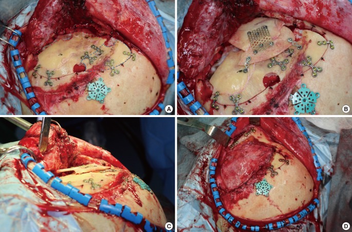
Fig. 2. Surgical detail.
(A) A pterional and temporal skull defect is demonstrated with a red dotted line corresponding to where the bone was removed during burr hole placement and pretemporal sphenoid wing drilling. The cranial flap was divided into two parts. The upper bone flap was bisected, and the outer table of this flap (blue dotted line) was used to close the rest of the craniotomy defect. The inner table of the upper bone flap was used as source material for the onlay graft. (B) A calvarial onlay graft was secured to the bone flap with titanium screws. (C) The onlay graft was made to overlap the cranial bone flap in order to ensure an appropriate thickness. Onlay grafts were prepared to overcorrect for soft tissue atrophy following the operation. (D) The temporalis muscle was reapproximated to the myofascial cuff along the superior temporal line.

