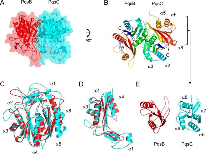
FIGURE 2.
Schematic diagrams of the PqsBCC129A structure. A, PqsBCC129A heterodimer is shown with PqsC (cyan) and PqsB (red) covered with a transparent molecular surface illustrating the interface. B, PqsB and PqsCC129A structures are colored in a rainbow from the N (blue) to the C terminus (red) with secondary structure elements labeled for PqsCC129A in all panels. C, superposition of PqsB and PqsCC129A. D, N-terminal sub-domains of PqsB(1–154) and PqsC(1–184) superposed with the catalytic dyad residue Ala-129 shown as sticks (purple). E, C-terminal sub-domains of PqsB(155–279) and PqsC(188–348) arranged side by side viewed with the same orientation showing PqsCC129A helix α6 is missing in PqsB. PqsC residue His-269 is shown as sticks (purple).

