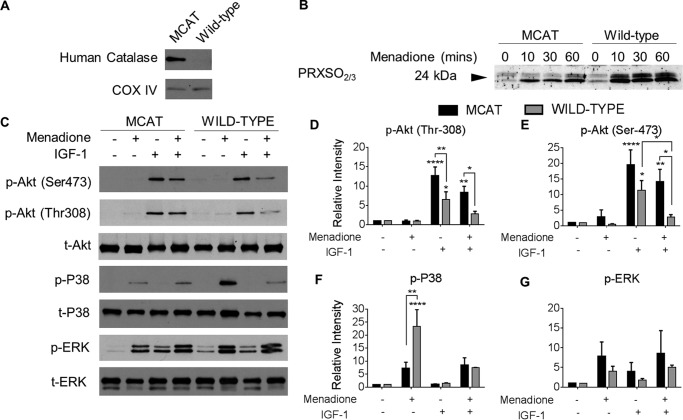
FIGURE 8.
Effect of oxidative stress on PRX hyperoxidation and kinase phosphorylation in chondrocytes from MCAT and wild-type mice. Femoral head cartilage explants from the hip joints of 4-week-old MCAT and wild-type mice were isolated and cultured in serum-supplemented medium for 48 h prior to overnight incubation in serum-free medium. Femoral head cartilage explants were treated with menadione (25 μm), IGF-1 (50 ng/ml), or combined treatments as indicated. Cell lysates were prepared by homogenization of explants as described under “Experimental Procedures.” A, cell lysates were immunoblotted for human catalase and for COX IV as a mitochondrial protein loading control. B, femoral cap cartilage explants from MCAT and wild-type mice were cultured in menadione for 0–60 min prior to incubation in NEM (100 mm, 10 min) to alkylate reduced thiols prior to lysis. Femoral caps were disrupted and homogenized in lysis buffer containing NEM. Hyperoxidized PRXs were detected by immunoblot using the PRX-SO2/3 antibody. C, for analysis of signaling, femoral cap cartilage explants were treated with menadione (30 min), IGF-1 (30 min), or pretreated with menadione (30 min) prior to IGF-1 treatment (30 min). Representative blots from n = 3 independent experiments are shown. D–G, results of densitometric analysis showing phosphorylation of Akt, p38, and ERK. Phosphorylation of proteins were normalized to total protein as a loading control and presented as fold change from untreated control. All data are presented as means ± S.E. from n = 3 independent experiments. Asterisks represent significant differences compared with control. *, p < 0.05; **, p < 0.01; ***, p < 0.001; ****, p < 0.0001 (ANOVA).

