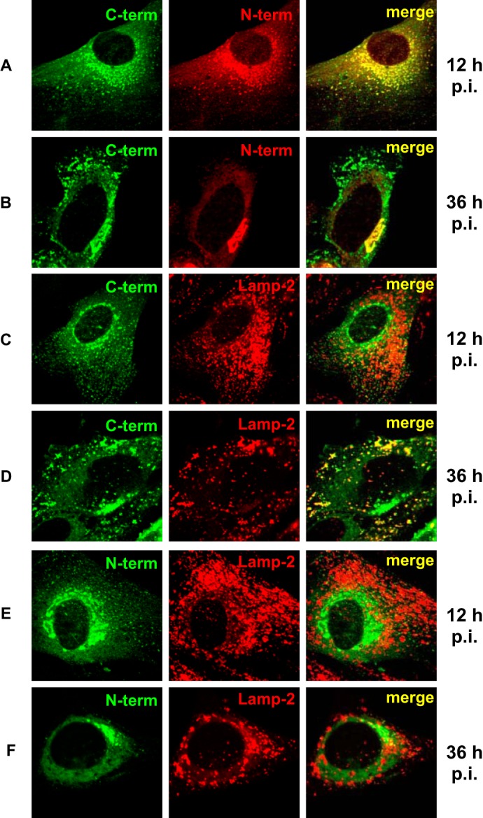
FIGURE 1.

Differential localization of the ectodomain and the cytoplasmic tail of E3/49K in the Golgi/TGN and late endosomes/lysosomes during early and late phases of infection. Primary fibroblasts (SeBu) were infected with Ad19a and processed for confocal laser microscopy at 12 or 36 h postinfection (p.i.), corresponding to the early and late phase of the infection cycle in these primary fibroblasts, respectively. The subcellular localization of E3/49K was analyzed with rabbit antiserum R25050 directed against the C terminus (C-term; A–D) and antiserum R48-7B (anti-49K-N; E and F), followed by FITC-labeled goat anti-rabbit IgG. For detection of the N-terminal part of E3/49K (N-term) the rat mAb 4D1 was employed (A and B, middle). This was compared with the late endosomal/lysosomal marker LAMP-2, using mouse mAb 2D5 (C and F, middle). Subsequent staining was done with donkey anti-rat IgG Texas Red and donkey anti-mouse IgG Rhodamine Red-X, respectively. One typical experiment of at least three is shown.
